In this virtual frog dissection, you will observe some of the external features and internal organs of a frog specimen. First, study each slide to begin making observations about the parts of a frog. You can click on each slide image to see a larger view.
The SkinThe first step is to place the specimen on the dissecting tray. 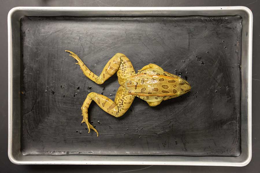
Dorsal Side 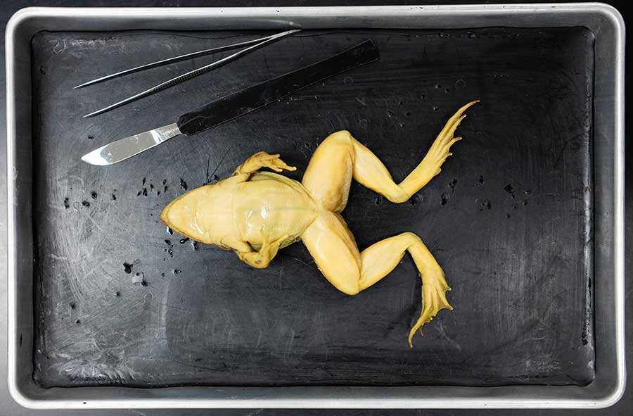
Ventral Side QuestionCompare the dorsal (back) and ventral (underneath) sides of the specimen. How does the skin on each side compare? The skin on the dorsal side is darker and has different colored lines and blotches. The ventral side skin is lighter and all one color.
The AppendagesThe next step is to study the appendages. 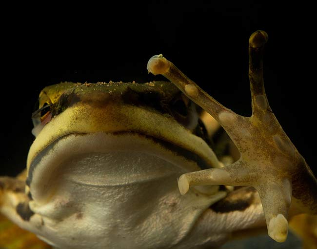
Front Limb 
Hindlimbs QuestionCompare the front limbs and the hindlimbs on the specimen. How many toes are on each? Are they webbed? The front limbs have 4 toes and are not webbed. The back limbs have 5 toes and are webbed.
The HeadThe next step is to study the external features of the head. 
Question
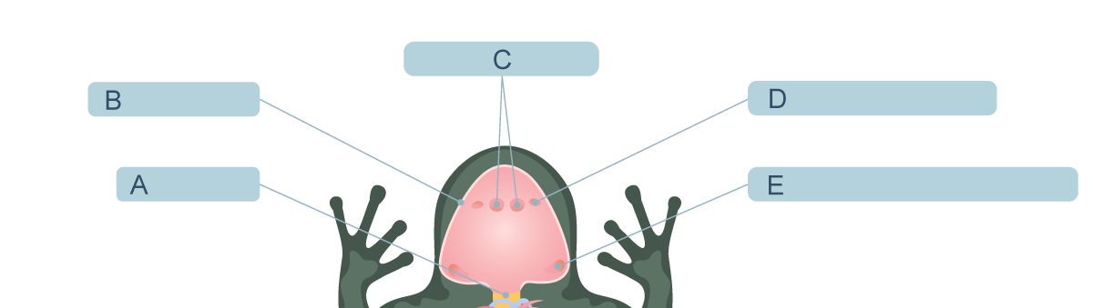
Locate the following parts on the frog head image below: tympanic membrane (ear) and nares (nostrils).

Inside of the HeadThe next step is to open the mouth wide and study the structures that are inside the head. View 1 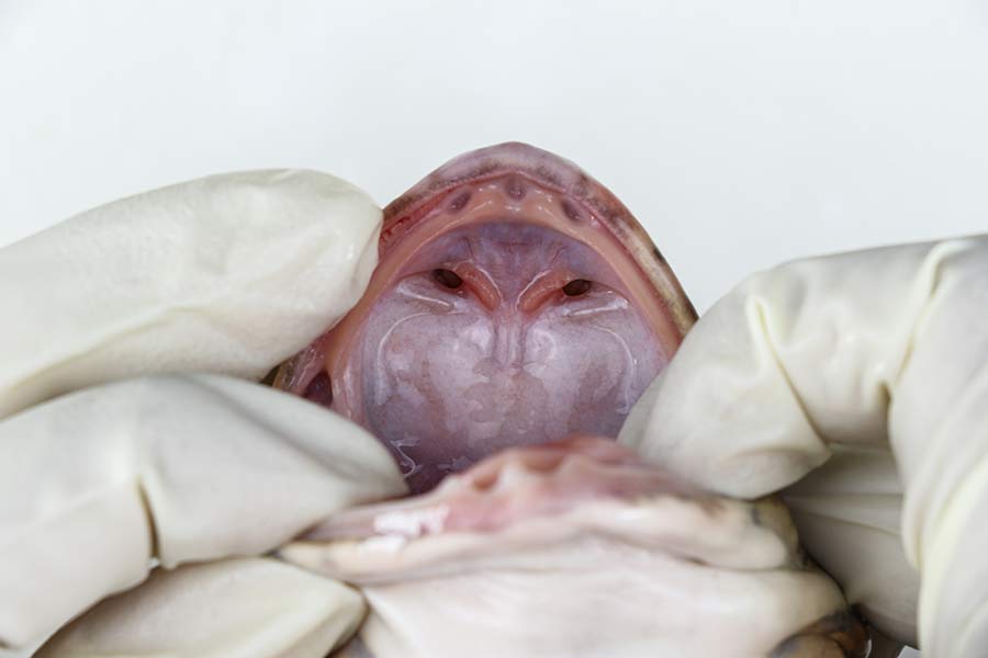
View 2 
Use the diagram to answer each question. Click the question to check your answer.
Under the SkinThe next step is to pin the limbs securely in the dissecting tray, with the ventral side facing upward. Then, make one longitudinal cut down the middle and two horizontal cuts near the arms. 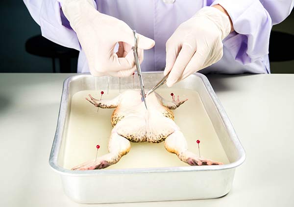
View 1 
View 2 QuestionAfter carefully cutting the skin, gently lift it back to expose the layer just underneath. Which layer is exposed just under the skin? A layer of muscle is exposed just under the skin.
Inside the BodyIn this step, cut the layer of muscle under the skin the same way you cut the skin and gently lift away this layer to reveal the inside of the body. 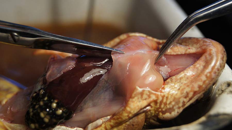
Cutting Open the Muscle 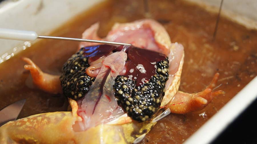
Lifting Back the Muscle QuestionThis specimen is female and is gravid with eggs. Where are the eggs located in the specimen? The eggs are in the lower sides of the abdominal cavity just under the muscle layer.
|
|