Let's investigate
In this virtual lab you will observe some of the internal organs of a frog specimen.
Before you begin, click the Activity button below to download your assignment worksheet. When you have completed the assignment, submit it to your teacher.
Locate the organs described and shown on each slide and label them on the diagram on your assignment worksheet.
|
After removing any eggs that are present, you can see the ventral organs. The heart is the small triangular organ in the upper central chest cavity, with vascular tissue connecting to it. The liver is the brown-colored organ and consists of three lobes. It is a large structure, filling a good portion of the body cavity and has many functions including removing toxins from the blood. The small intestine is the long tube that leads from the stomach. The small intestine is surrounded by connective tissue with many blood vessels for nutrient absorption. 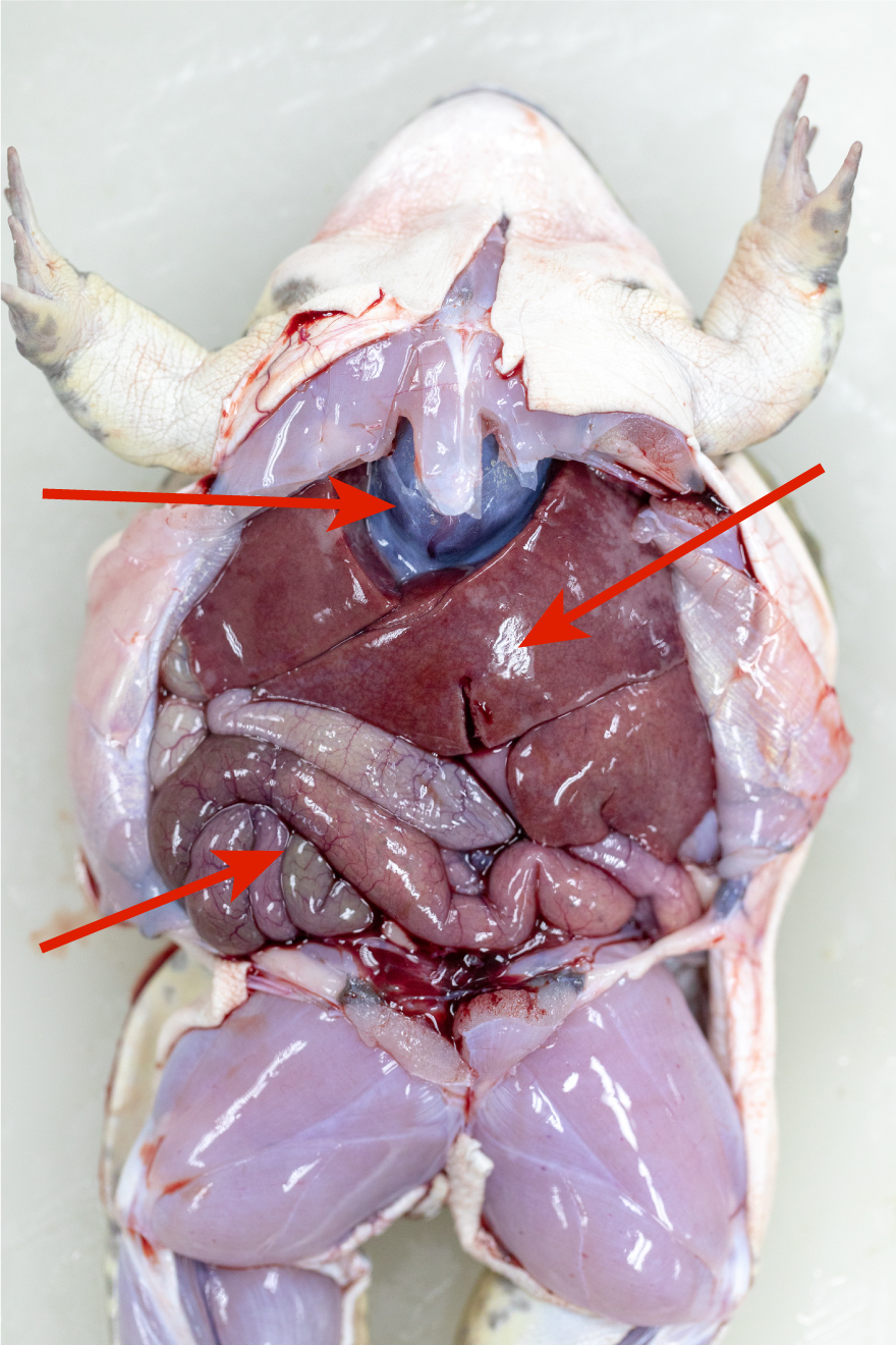
Three organs are visible on the specimen on this slide. Locate them, then label them on the diagram on your assignment worksheet. Moving the ventral organs aside slightly helps to reveal more organs underneath them. The gall bladder is a small, greenish-brown sac that stores bile made in the liver. Curving from underneath the liver is the stomach, which holds food that enters the body before it travels through the intestines. 
Two more organs are visible on the specimen on this slide. Locate them, then label them on the diagram on your assignment worksheet. Because the body cavity of a frog is 3-dimensional, moving organs slightly helps reveal others that are hidden. As you follow the small intestine down, it widens into the large intestine, which is where water and salts are absorbed from waste The large intestine leads to the cloaca, which is a cavity where urine, sperm, eggs, and feces exit the body. 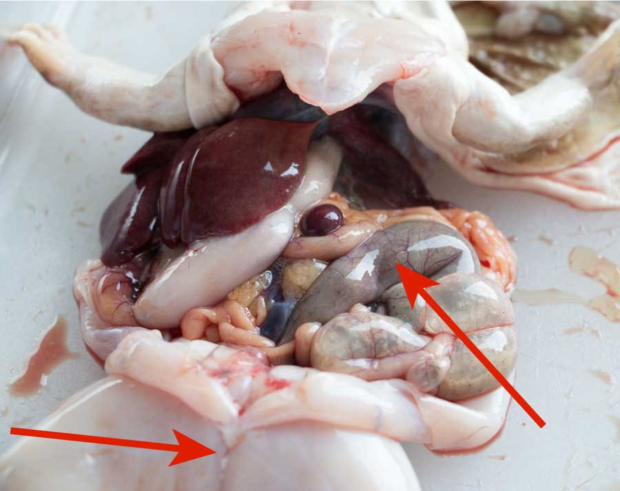
Two more features are visible on the specimen on this slide. Locate them, then label them on the diagram on your assignment worksheet. Once many of the internal organs in the body cavity are removed, another pair of organs is visible. The lungs are located on either side of the heart and are made of a spongy tissue. 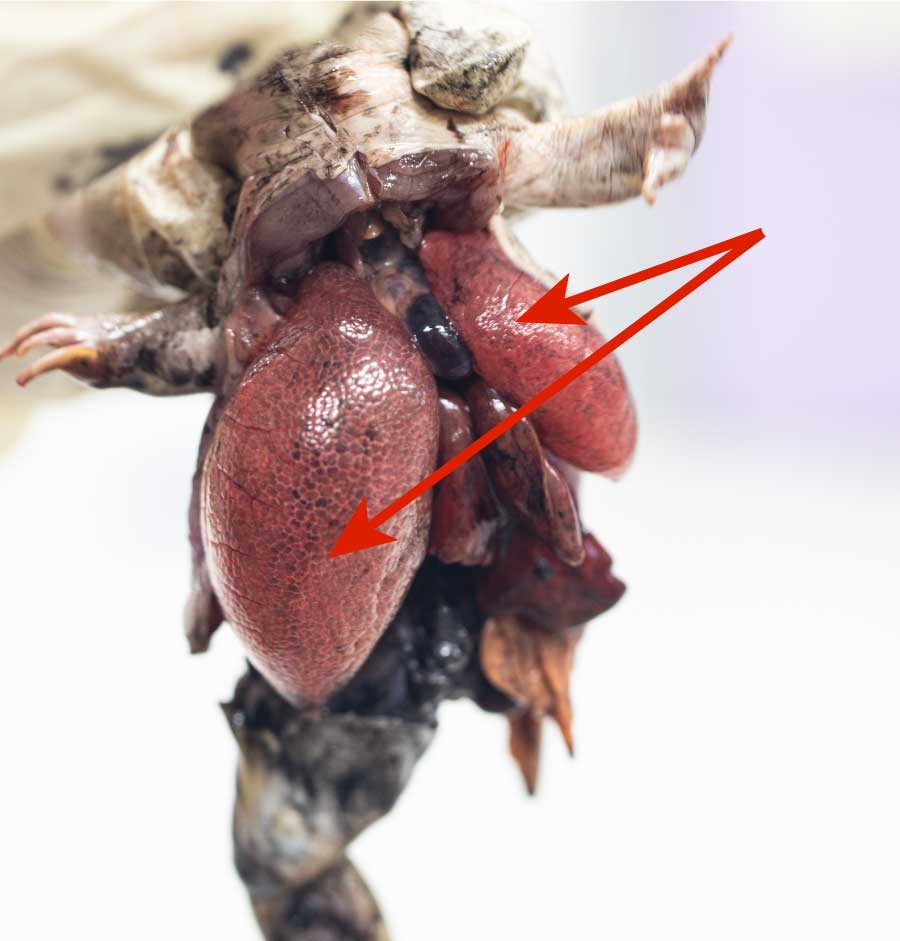
A pair of organs is visible on the specimen on this slide. Locate them, then label them on the diagram on your assignment worksheet. |
|