The video on the previous page shows the entire process of mitosis from start to finish. To understand upcoming lessons in this course, you'll need to know some key details about each phase of mitosis. Use this interactive table to focus on each phase--click the phase to see what details are important to remember.
| Prophase | In this phase the chromosomes have condensed and can be seen through a light microscope. Sister chromatids will also become visible in the [X] formation. The mitotic spindle begins to form as the centrosomes, which produce the spindles, move away from each other. | 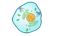 |
| Prometaphase | In this phase the nuclear envelope begins to break down. The microtubule spindle fibers emerging from the centrosomes begin to enter the nuclear area of the cell. The sister chromatids can be seen clearly. They are attached to the spindle fibers and begin to jerk back and forth. | 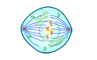 |
| Metaphase | This phase lasts longer than the other mitotic phases, about 20 minutes. During the metaphase, centromeres, which are at the opposite ends of the cell, use spindle fibers to place the chromosomes in the proper place in the cell. | 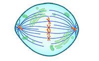 |
| Anaphase | This phase is the shortest, lasting only two minutes. During this time, the sister chromatids begin to separate, moving towards opposite ends of the cell as the microtubular spindle fibers shorten. (Think of line dancers letting go of their partner's hands and moving away from each other.) After they separate, the chromatids are full chromosomes. At the end of this phase, the two ends of the cell have an equal number of chromosomes. In case of the cheek cells, there are 46 chromosomes at each end. | 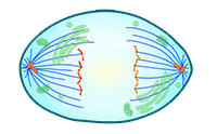 |
| Telophase | In this phase, two nuclei develop and encompass the chromosomes at each end of the cell. At this point, the cell looks like an egg with two yolks. The cell has two complete nuclei, and mitosis is complete. | 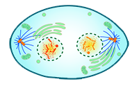 |
| Cytokinesis | By the end of telophase, the cytoplasm has begun to divide. In animals cells, like the cheek cells you have been studying, a cleavage furrow forms and pinches the cell into two daughter cells. In plants cells, like those in an onion root tip, a cell plate forms. | 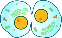 |
Question
Why would the cells at the tips of plant roots, such as the roots of an onion, work well for studying mitosis?
Roots grow from the tips, so you can be sure that there will be cells in the process of dividing there.