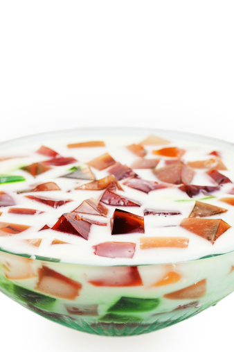
 Sponges are multicellular, asymmetrical, sessile animals. They are the simplest kind of animal because they lack organs and are the only animals that lack tissue. Sponges are essentially a clump of cells suspended within a jelly-like substance called mesohyl that holds all the cells together.
In the dessert pictured, if the sponge cells are represented by the colorful cubes, which part would represent the mesohyl? Think of the answer, then click "Show Me" to see if you are correct.

The cream that the cubes are floating in represents the mesohyl.
Even though sponges lack tissue, they are certainly not without their own level of complexity and organization. In fact, sponges lay the framework for all animal bodies, in that all the animals that have come after sponges are designed similarly but have become ever so slightly more complex over time.
There are different types of cells within a sponge that have a loose coordination with each other and that can even recognize each other. For example, if you to cut apart a sponge, the pieces would recognize each other as "self" and recombine, or, even more spectacularly, each piece can grow into a new copy the original sponge.
In the activity below, click each tab to read about how a sponge is organized and the different types of cells found within a sponge.
Ostia
Choanocytes
Amoebocytes
Oscula
Epidermal Cells
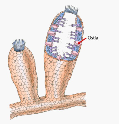
There are many tiny openings, or pores, on the surface of all sponges. They are shaped like little tunnels, which can be seen in the sponge pictured. It's these pores that give the sponge phylum the name Porifera. These pores are called ostia.
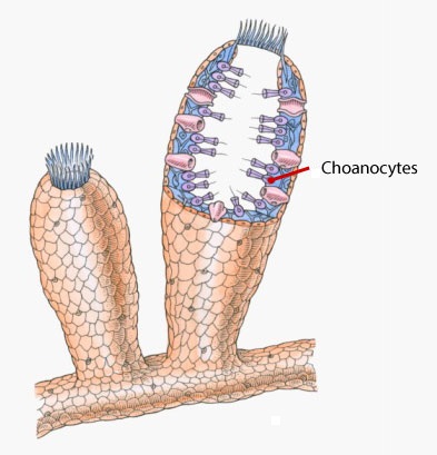
Choanocytes line the wall of the internal cavity of the sponge, as indicated on the picture. They are flagellated (have flagella) and are called collar cells because of what appears to be a collar surrounding the flagellum. What animal-like protist do these choanocytes resemble? Think of the answer, then click "Show Me" to see if you are correct.

Choanocytes resemble the protist choanoflagellate.
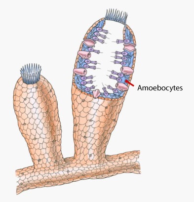
Amoebocytes are amoeba-like cells that are capable of moving throughout the mesohyl, which is the gel-like matrix that holds all the sponge cells together. They are the blue cells in the image and are sandwiched in between the inner and outer surfaces of the sponge.
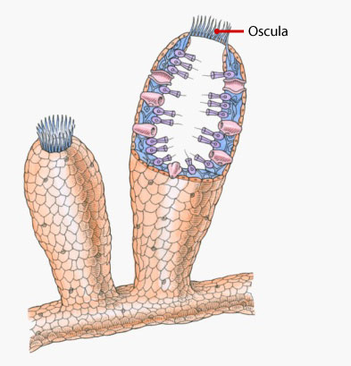
One end of a sponge attaches to hard surface like a rock or a coral, while the other end has large openings, called oscula (singular: oscula).
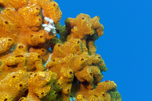 All the many openings visible on the sponge pictured are the oscula. Note that in the first image, there are only two oscula, located on the very top of the sponge.
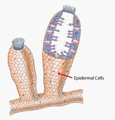
The outer surface of the sponge is covered in a layer of thin, flattened cells, which are the epidermal cells of the sponge. The epidermal cells are tan colored in the picture.
In the activity below, find the definition on the right side that corresponds with a word on the left. Make your selection by dragging a circle on the left to its match on the right.
|
epidermal cells
amoebocytes
choanocytes
osculum
ostia
mesohyl
|
large opening in sponges
gel-like matrix that sponge cells are suspended within
flagellated cells that line the inner cavity of sponges
flattened cells lining the outer surface of the sponge
tiny openings on the surface of the sponge
cells that move through the mesohyl of a sponge
|
Question
Scientists believe that about 700 million years ago, choanoflagellates and amoebas (which are unicellular) aggregated to form the first primitive animal--a sponge. What do you think are some advantages to being multicellular as opposed to unicellular?
